Zygote Drawing
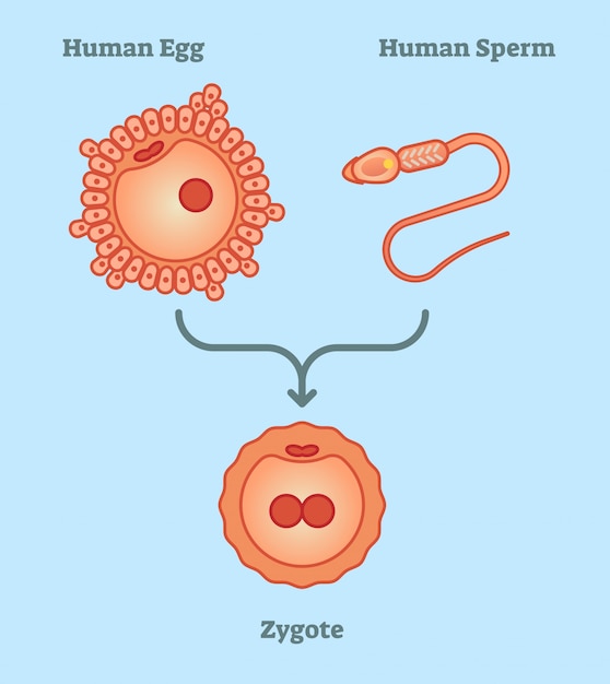
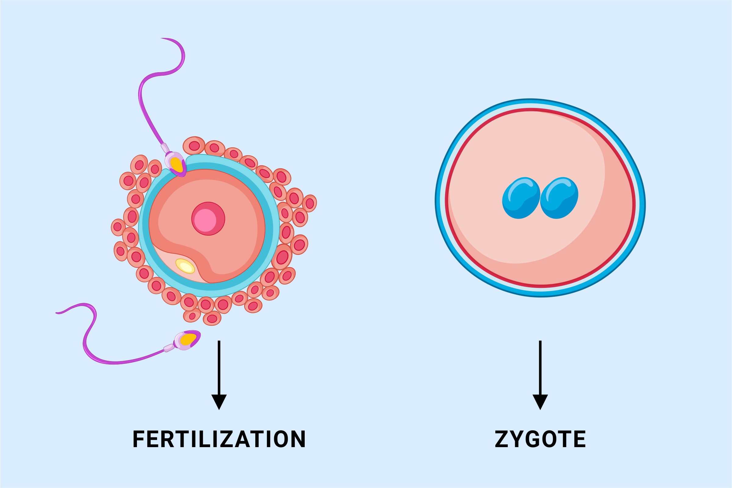
Zygote Drawing - Your 3d graphics will be artistic and accurate with the zygote's amazing blend of medical precision and high aesthetic quality. The zygote stage development occurs in the first week of fertilization. Gametes have half the chromosomes (haploid) of a typical body cell, while zygotes have the full set (diploid). Department of defense and nasa. One fuses with the egg cell to form zygote and the other fuses with the polar nuclei to form the endosperm (other seed parts) Well, this right over here is a picture of a zygote which is this fertilized, you could view it as a fertilized egg, it's now starting to have all 46 chromosomes, and what's interesting here is you can see the nuclei from the two cells, they haven't completely fused yet. Embryo development 19 century medical illustration The genome of the zygote is the combination of dna in each gamete and contains all the genetic information required to form an individual. The fusion is known as fertilization. The zygote's genome is a combination of the dna in each gamete, and contains all of the genetic information of a new. The dna material from the two cells is combined in the resulting zygote. Web a zygote is the first diploid cell that is formed by the fusion of male and female gametes resulting in the formation of an embryo. Web a zygote is the cell formed when two gametes fuse during fertilization. Photograph of the original illustration from a system. Web your first form as a zygote split to make two cells. Web sperm and egg cells, known as gametes, fuse during fertilization to create a zygote. Newest results embryo development 19 century medical illustration photograph of the original illustration from a system of human anatomy by erasmus wilson published in 1859. There are two ways cell division can happen. Gametes have half the chromosomes (haploid) of a typical body cell, while zygotes have the full set (diploid). We would be happy to talk about our process and demo the models for further clarification. A zygote is the single cell formed when an egg and a sperm cell fuse; Web but what does it look like as soon as fertilization. Most popular abstract doodle pattern with microbe, virus, bacteria. Our solid models have no parallel on the market and have been used in multiple projects by both the u.s. Web but what does it look like as soon as fertilization has occurred? Web a zygote is the first diploid cell that is formed by the fusion of male and female. Department of defense and nasa. The snapshot icon at the top center will take a snapshot of your scene that can then be saved as a jpg or drawn on with the included pen tools. Web browse 30+ human zygote drawing stock photos and images available, or start a new search to explore more stock photos and images. The genome. Web browse 30+ human zygote drawing stock photos and images available, or start a new search to explore more stock photos and images. The zygote's genome is a combination of the dna in each gamete, and contains all of the genetic information of a new. Web but what does it look like as soon as fertilization has occurred? A zygote. Most popular abstract doodle pattern with microbe, virus, bacteria. Web but what does it look like as soon as fertilization has occurred? We would be happy to talk about our process and demo the models for further clarification. Web there are 2 male gametes in a pollen grain. Web a zygote is the first diploid cell that is formed by. Well, this right over here is a picture of a zygote which is this fertilized, you could view it as a fertilized egg, it's now starting to have all 46 chromosomes, and what's interesting here is you can see the nuclei from the two cells, they haven't completely fused yet. The zygote’s first priority is dividing to make lots of. Web zygote, fertilized egg cell that results from the union of a female gamete (egg, or ovum) with a male gamete. Gametes have half the chromosomes (haploid) of a typical body cell, while zygotes have the full set (diploid). Zygote body is a free online 3d anatomy atlas. The snapshot icon at the top center will take a snapshot of. Then those cells split, making four.and so on and so forth, until you became the living, functioning organism you are today. Web zygote's cad models are created using actual medical scan data from 50th percentile male and females. We would be happy to talk about our process and demo the models for further clarification. Web browse 30+ human zygote drawing. Web but what does it look like as soon as fertilization has occurred? Web hi friends, in this video we will learn to draw draw formation and development of an embryo from the zygote class 8 (this diagram is based on c. Your 3d graphics will be artistic and accurate with the zygote's amazing blend of medical precision and high aesthetic quality. Learn how a zygote, the single cell produced by fertilization, divides by mitosis to produce all the tissues of the human body (including germ cells, which can undergo meiosis to make sperm and eggs). The zygote’s first priority is dividing to make lots of new cells, so its first few days are spent in rapid mitotic. The cellular mechanisms present in the gametes also function in the zygote, but the newly fused dna produces a different effect in the new cell. Hello friends welcome to my. Zygote body is a free online 3d anatomy atlas. The dna material from the two cells is combined in the resulting zygote. Web your first form as a zygote split to make two cells. Embryo development 19 century medical illustration The zygote's genome is a combination of the dna in each gamete, and contains all of the genetic information of a new. Web sperm and egg cells, known as gametes, fuse during fertilization to create a zygote. Gametes have half the chromosomes (haploid) of a typical body cell, while zygotes have the full set (diploid). Newest results embryo development 19 century medical illustration photograph of the original illustration from a system of human anatomy by erasmus wilson published in 1859. Web hi friends, in this video we will learn how to draw diagram of fertilisation in plants〰️〰️〰️〰️〰️〰️〰️〰️〰️〰️〰️〰️〰️〰️〰️〰️〰️👉.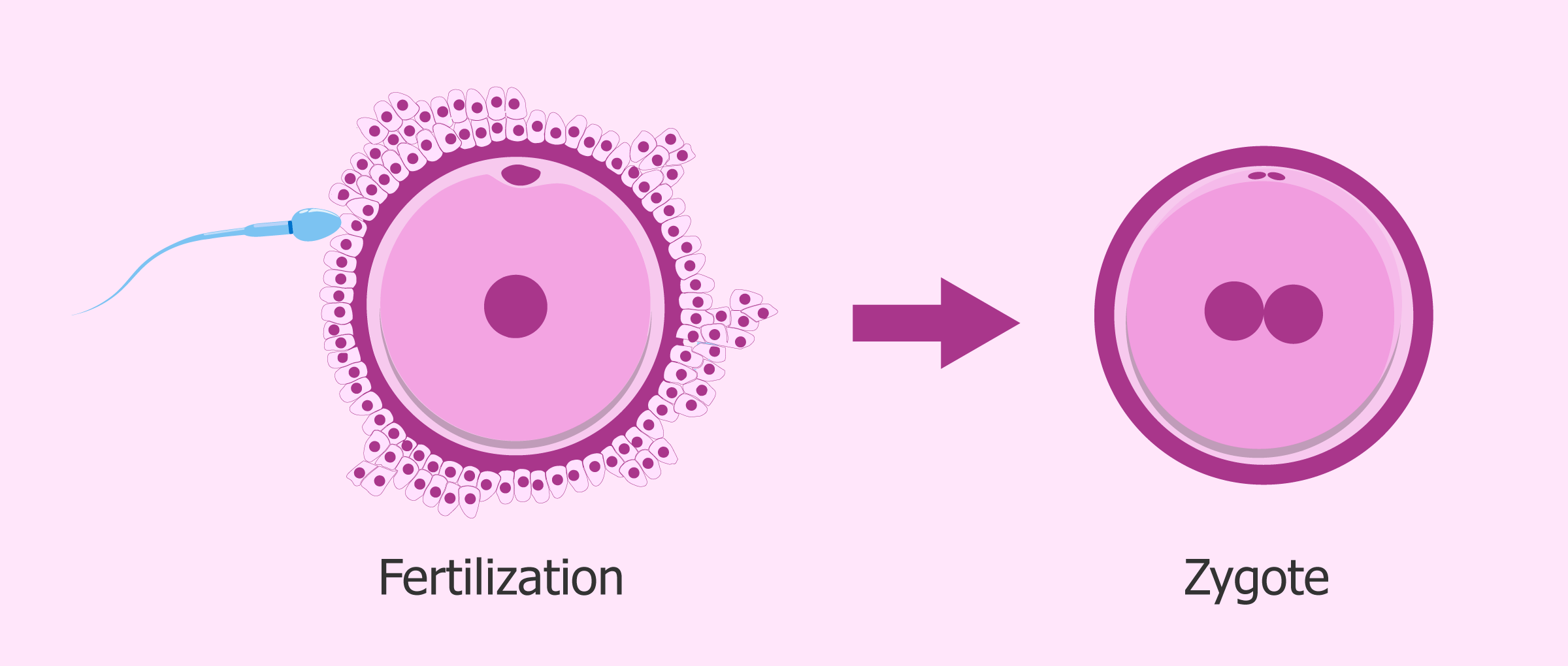
Zygote Definition, Formation and Examples
Zygote Vectoriels et illustrations libres de droits iStock
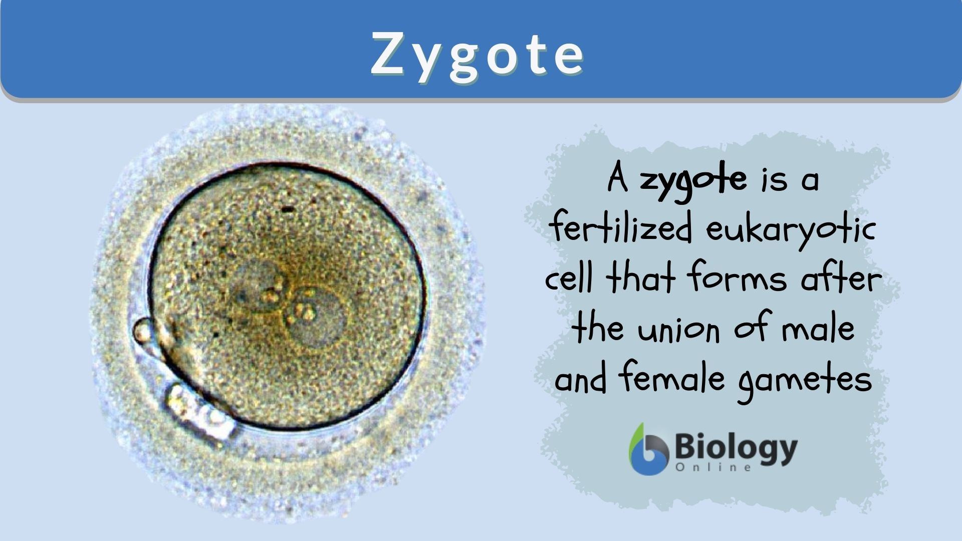
Zygote Definition and Examples Biology Online Dictionary
Premium Vector What is zygote vector illustration diagram, simple
What is a Zygote in biology? Biology concepts Biology questions

30+ Human Zygote Drawing Stock Photos, Pictures & RoyaltyFree Images
Best Human Zygote Illustrations, RoyaltyFree Vector Graphics & Clip
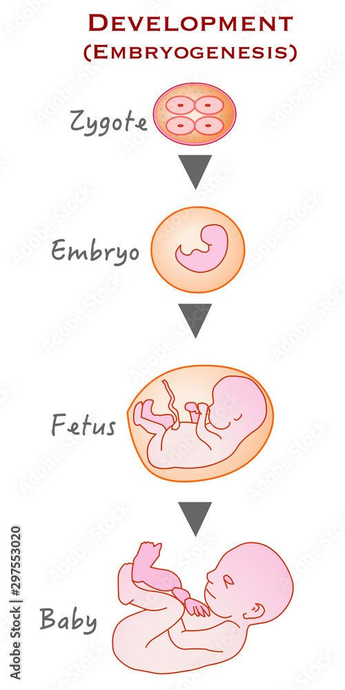
From zygote to infant. Formation, development and growth stages. Zygote
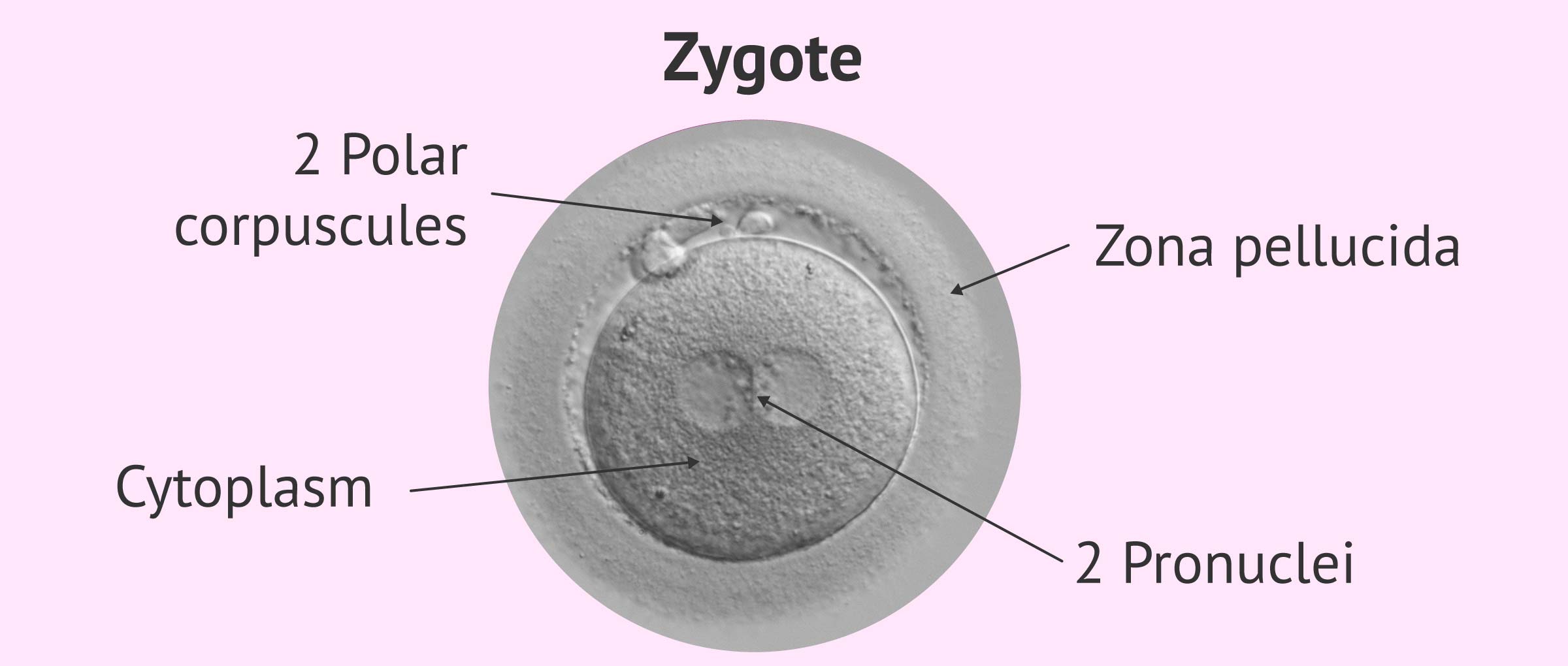
Structure of a zygote

how to draw or sketch art zygote of human Art sketches, Pictures to
Web A Zygote Is The First Diploid Cell That Is Formed By The Fusion Of Male And Female Gametes Resulting In The Formation Of An Embryo.
Our Solid Models Have No Parallel On The Market And Have Been Used In Multiple Projects By Both The U.s.
A Zygote Is The Single Cell Formed When An Egg And A Sperm Cell Fuse;
From Ancient Greek Ζυγωτός (Zygōtós) 'Joined, Yoked', From Ζυγοῦν (Zygoun) 'To Join, To Yoke') Is A Eukaryotic Cell Formed By A Fertilization Event Between Two Gametes.
Related Post:

