Ultrasound Drawing
Ultrasound Drawing - Free for commercial use high quality images Web a step by step guide to a basic ultrasound protocol to image the liver. Web with deeper structures, other tissues and densities (eg, bone, gas) can interfere with images. Web begin by scanning large areas (sometimes called survey views) and then focus in to smaller areas of interest. Web find & download free graphic resources for ultrasound medical. For example, to detect valvular and chamber size abnormalities and to estimate ejection fraction and myocardial strain (see echocardiography. Learn how to draw ultrasound pictures using these outlines or. Web an ultrasound probe actually contains an array of multiple tiny component piezoelectric transducers each providing a single line of ultrasound data. The images can provide valuable information for diagnosing and directing treatment for a variety of diseases and conditions. Here presented 53+ ultrasound drawing images for free to download, print or share. 99,000+ vectors, stock photos & psd files. For most humans audible sound ranges between 20 hz and 20,000 hz (20 khz) ultrasound refers to any sound waves with frequencies greater than 20khz. For example, to detect valvular and chamber size abnormalities and to estimate ejection fraction and myocardial strain (see echocardiography. Medical therapy surgery vector sketch equipment. The illustration shows. Web begin by scanning large areas (sometimes called survey views) and then focus in to smaller areas of interest. Ultrasound imaging is rapidly replacing radiological methods in the investigation of pelvic floor disorders. Web browse 820+ ultrasound drawings stock photos and images available, or start a new search to explore more stock photos and images. Web a step by step. Compared to the previous blind venipuncture, used mainly in the 80s and 90s, the positioning with ultrasound probe has demonstrated concrete advantages in terms of: Medical surgery or therapy equipment sketch icons. Learn how to draw ultrasound pictures using these outlines or. Increased percentage of successes at first attempt in. Web welcome to ultrasound 101. Transrectal, transvaginal/introital and transperineal/translabial methods are being employed, with the latter probably the most widespread due to ease of use and availability of equipment. 99,000+ vectors, stock photos & psd files. These transducers are optimal for examining larger organs from between the ribs. Web this article provides a beginners guide to ultrasound (pocus), including how ultrasound works and how ultrasound. You can find more ultrasound drawing in our search box. Web with deeper structures, other tissues and densities (eg, bone, gas) can interfere with images. The images can provide valuable information for diagnosing and directing treatment for a variety of diseases and conditions. Ultrasound imaging is rapidly replacing radiological methods in the investigation of pelvic floor disorders. Arbitrary section planes. You can find more ultrasound drawing in our search box. Increased percentage of successes at first attempt in. Medical therapy surgery vector sketch equipment. Web we have collected 36+ original and carefully picked ultrasound drawing in one place. Web choose from 600 drawing of the ultrasound stock illustrations from istock. Feel free to download, share and use them! Medical therapy surgery vector sketch equipment. The illustration shows a schematic drawing of wave length, pressure and amplitude. Transrectal, transvaginal/introital and transperineal/translabial methods are being employed, with the latter probably the most widespread due to ease of use and availability of equipment. Web are you looking for the best images of ultrasound. Web welcome to ultrasound 101. You can find more ultrasound drawing in our search box. Web with deeper structures, other tissues and densities (eg, bone, gas) can interfere with images. Web begin by scanning large areas (sometimes called survey views) and then focus in to smaller areas of interest. Transrectal, transvaginal/introital and transperineal/translabial methods are being employed, with the latter. Web are you looking for the best images of ultrasound drawing? Web begin by scanning large areas (sometimes called survey views) and then focus in to smaller areas of interest. Medical therapy surgery vector sketch equipment. Web welcome to ultrasound 101. Arbitrary section planes and “virtual travels through the body” are possible. By seamlessly stitching together each of these lines of data, the display creates a 2d image in the same way that old television screens created images from parallel lines of display. You can find more ultrasound drawing in our search box. Ultrasonography is commonly used to evaluate the following: Geared to the new sonographer student, major landmarks and tips and. Web this article provides a beginners guide to ultrasound (pocus), including how ultrasound works and how ultrasound can be used in clinical practice. You can find more ultrasound drawing in our search box. For example, to detect valvular and chamber size abnormalities and to estimate ejection fraction and myocardial strain (see echocardiography. Web choose from 600 drawing of the ultrasound stock illustrations from istock. Learn how to draw ultrasound pictures using these outlines or. Geared to the new sonographer student, major landmarks and tips and tricks are covered while drawing the common images. Web are you looking for the best images of ultrasound drawing? Web we have collected 36+ original and carefully picked ultrasound drawing in one place. Web begin by scanning large areas (sometimes called survey views) and then focus in to smaller areas of interest. The images can provide valuable information for diagnosing and directing treatment for a variety of diseases and conditions. By seamlessly stitching together each of these lines of data, the display creates a 2d image in the same way that old television screens created images from parallel lines of display. Web basic transducer formats are sector, linear array, and curved array ( fig. Ultrasound imaging is rapidly replacing radiological methods in the investigation of pelvic floor disorders. Web a step by step guide to a basic ultrasound protocol to image the liver. Web diagnostic ultrasound, also called sonography or diagnostic medical sonography, is an imaging method that uses sound waves to produce images of structures within your body. Arbitrary section planes and “virtual travels through the body” are possible.
Ultrasound image and drawing to demonstrate the fetal head direction
Fetal ultrasound hand drawn outline doodle icon. Pregnancy sonogram of
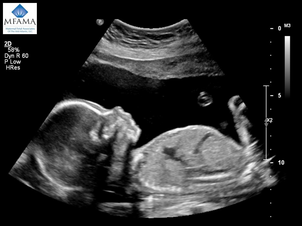
Anatomy Ultrasound Maternal Fetal Associates of the MidAtlantic
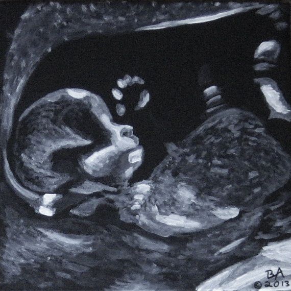
Ultrasound Drawing at GetDrawings Free download
Baby Ultrasound Art Watercolor Sonogram Print Baby Shower Etsy

Illustration of a Woman Undergoing a Chest Ultrasound Stock Vector
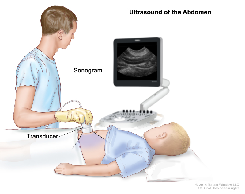
Ultrasound Drawing at GetDrawings Free download
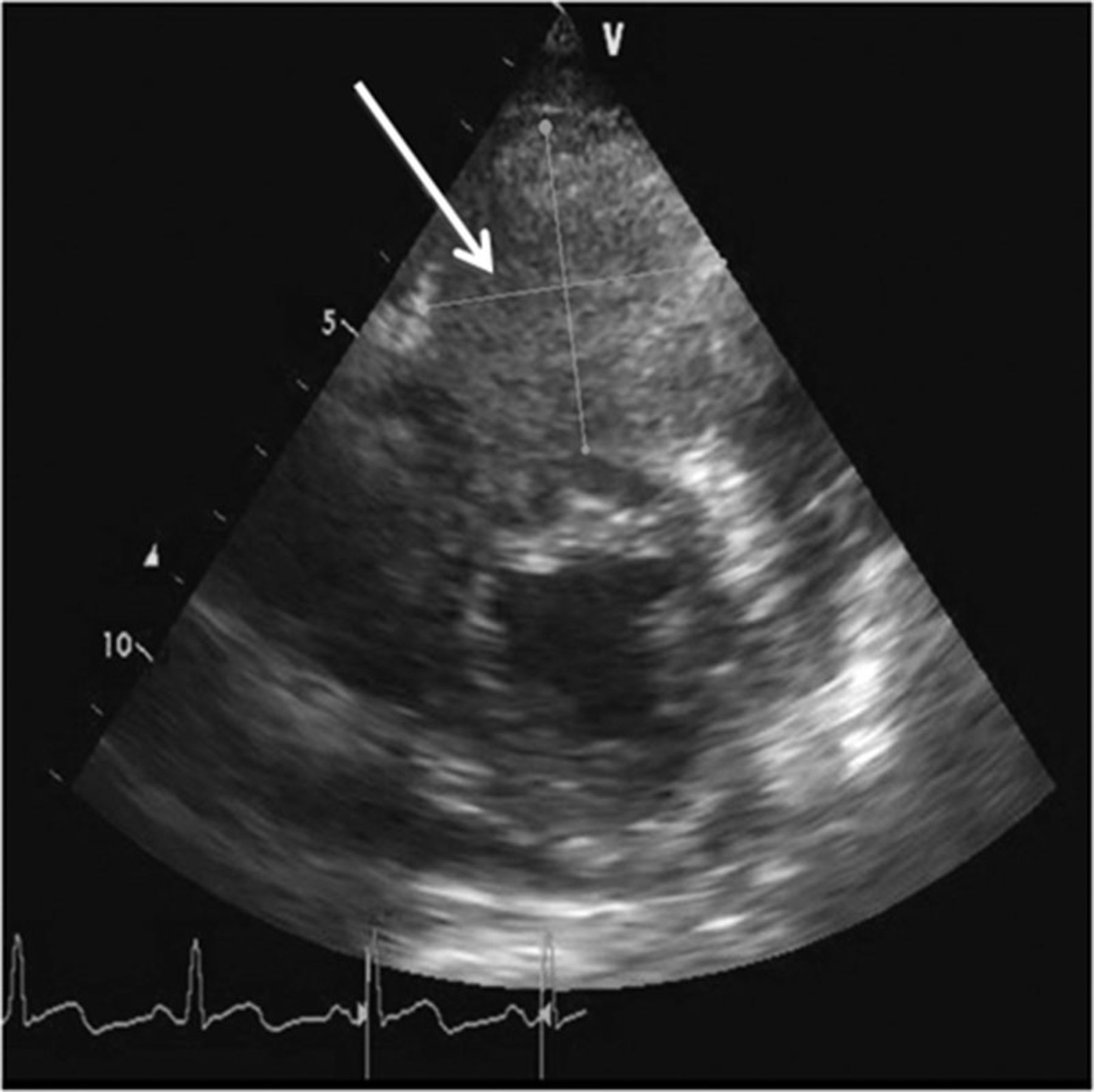
Ultrasound Drawing at GetDrawings Free download

ultrasound scan of baby 2610654 Vector Art at Vecteezy
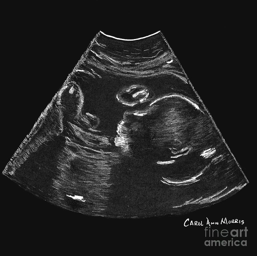
Ultrasound baby Drawing by Carol Morris Fine Art America
Compared To The Previous Blind Venipuncture, Used Mainly In The 80S And 90S, The Positioning With Ultrasound Probe Has Demonstrated Concrete Advantages In Terms Of:
Web Find & Download Free Graphic Resources For Ultrasound Medical.
Web Pencil Sketch Ultrasound Portrait, Baby Scan Keepsake, Family Portrait, Personalized Sonogram Drawing From Photo, Pregnancy Digital Drawing.
These Transducers Are Optimal For Examining Larger Organs From Between The Ribs.
Related Post: